
Micro-CT render of the cortical bone in a rat tibia
This image shows a render of the canal network and some larger osteocyte lacunae in a rat tibia. It was taken using a SkyScan desktop micro-CT. Most of my research has focused on using micro-CT to image cortical bone.

Radially orientated vascular canals in the humeral cortical bone of a red-tailed hawk.
The main question I focused on for my PhD was investigating the relationship between vascular canal orientation and flight and growth rate. This study looked at the differences in orientation between humeri and femora in birds and bats.
Interpreting the three‐dimensional orientation of vascular canals and cross‐sectional geometry of cortical bone in birds and bats. 2018. IV Pratt, JD Johnston, E Walker, DML Cooper. Journal of anatomy 232 (6), 931-942
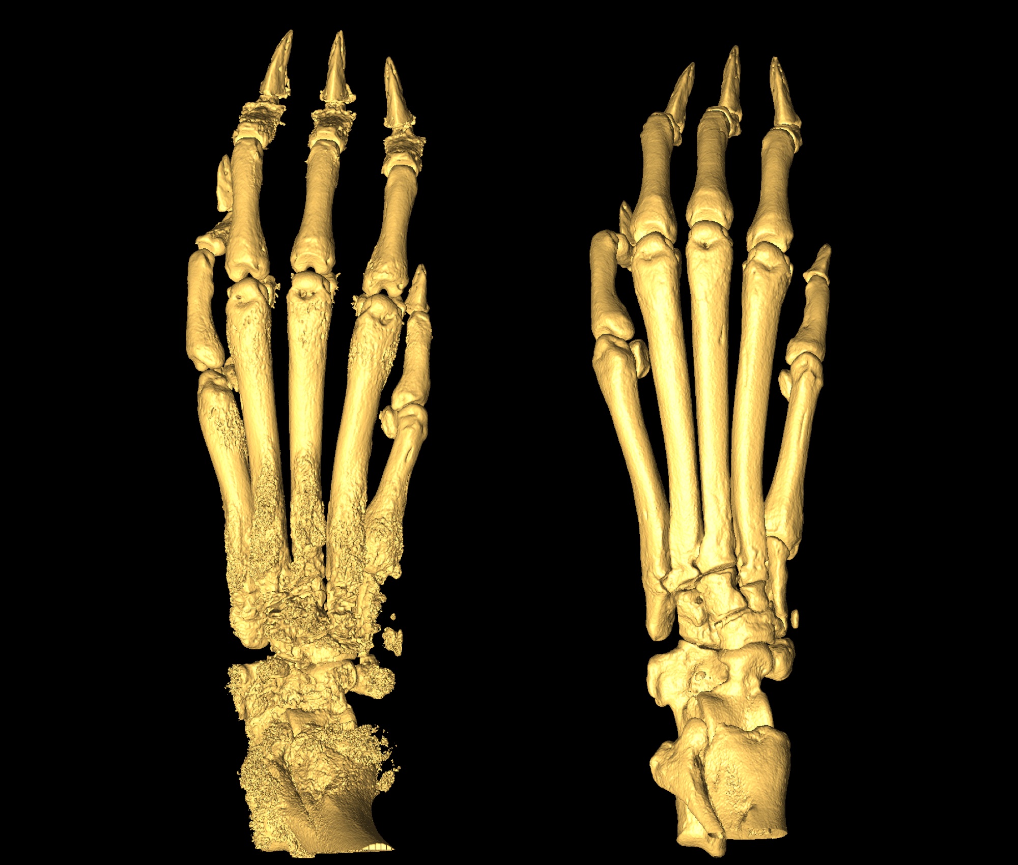
Metacarpal joints in arthritic rats
One of my earliest projects was imaging rat paws for a project describing a collgen-induced arthritis model in rats. The right image shows a normal non-arthritic paw, and the left shows an induced arthritis condition. Scans were taken using a SkyScan 1172 desktrop micro-CT and surface renders were created in Amira.
Age-related differences in collagen-induced arthritis: clinical and imaging correlations. 2013. TD Wilson-Gerwing, IV Pratt, DML Cooper, TI Silver, AM Rosenberg. Comparative medicine 63 (6), 498-502

Micro-CT slices of broiler chicken humeri and femora
Following on the research path looking at vascular canal orientation, I ran a project investigating the effect of growth rate on canal orientation in broiler chickens. This project was a challenge for me to co-ordinate a lot of moving pieces. Working with live animals and advanced synchrotron imaging, I relied of a lot of different people for their expertise to bring everything together.

Eocene insect trapped in amber
Using micro-CT we can image ancient creatures trapped in amber and reveal their three dimensional structure. In this image both the external and internal structure are revealed. This work was done in collaboration with researchers at the Royal Saskatchewan Museum.

Scanning Electron Microscopy image of human femoral cortical bone
Scanning electron microscopy is another technique that can be used to image bone at extremely high resolution. This image was taken for a project looking at the branching of cortical canals.

Histological image of cortical bone in a Hadrosaur tibia
While I believe micro-CT represents the future of bone microstructure imaging, traditional techniques like histological imaging still offer very high resolution details, although it comes at the cost of only imaging in two dimensions.

Bronze age siberian mandible with embedded projectile point
Micro-CT is a great technique for its ability to reveal the inner details of structures. This project investigated a bronze age siberian mandible that had an embedded projectile point. We used several different CT modalities to investigate the effects of the arrow head on the bone. Using synchrotron micro-CT, we were able to reveal the details of the microstructure and determine that no remodeling or bony repair had taken place after the injury. This meant that this injury occured at or around the time of death of the individual.
Point taken: an unusual case of incisor agenesis and mandibular trauma in Early Bronze Age Siberia. 2014. AR Lieverse, IV Pratt, RJ Schulting, DML Cooper, VI Bazaliiskii, AW Weber. International Journal of Paleopathology 6, 53-59
Interpreting the three‐dimensional orientation of vascular canals and cross‐sectional geometry of cortical bone in birds and bats. 2018. IV Pratt, JD Johnston, E Walker, DML Cooper. Journal of anatomy 232 (6), 931-942
Occurrence of osteon banding in adult human cortical bone. 2017. JM Andronowski, IV Pratt, DML Cooper. American journal of physical anthropology 164 (3), 635-642
Evaluating differential nuclear DNA yield rates and osteocyte numbers among human bone tissue types: A synchrotron radiation micro-CT approach. 2017. JM Andronowski, AZ Mundorff, IV Pratt, JM Davoren, DML Cooper. Forensic Science International: Genetics 28, 211-218
A method for measuring the three-dimensional orientation of cortical canals with implications for comparative analysis of bone microstructure in vertebrates. 2017. IV Pratt, DML Cooper. Micron 92, 32-38
In vivo imaging of rat cortical bone porosity by synchrotron phase contrast micro computed tomography. 2014. IV Pratt, G Belev, N Zhu, LD Chapman, DML Cooper. Physics in Medicine & Biology 60 (1), 211
Point taken: an unusual case of incisor agenesis and mandibular trauma in Early Bronze Age Siberia. 2014. AR Lieverse, IV Pratt, RJ Schulting, DML Cooper, VI Bazaliiskii, AW Weber. International Journal of Paleopathology 6, 53-59
Age-related differences in collagen-induced arthritis: clinical and imaging correlations. 2013. TD Wilson-Gerwing, IV Pratt, DML Cooper, TI Silver, AM Rosenberg. Comparative medicine 63 (6), 498-502
Improved compressed sensing-based algorithm for sparse-view CT image reconstruction. 2013. Z Zhu, K Wahid, P Babyn, D Cooper, I Pratt, Y Carter. Computational and mathematical methods in medicine 2013